Roof of this chamber contains the bicuspid valve – The bicuspid valve, located in the heart’s left atrium, plays a crucial role in regulating blood flow. This article delves into the anatomy, function, and clinical significance of the bicuspid valve, exploring its structural components, mechanism of action, and implications for cardiovascular health.
The bicuspid valve consists of two leaflets, chordae tendineae, and papillary muscles. During the cardiac cycle, the valve opens and closes to allow blood to flow from the left atrium to the left ventricle.
Anatomical Location and Structure: Roof Of This Chamber Contains The Bicuspid Valve

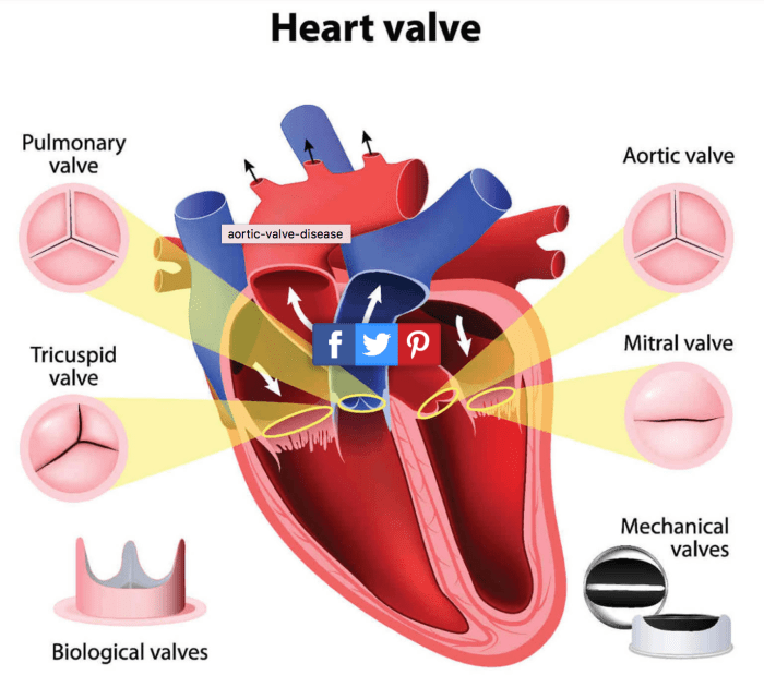
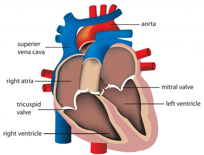
The bicuspid valve, also known as the mitral valve, is located in the heart chamber between the left atrium and left ventricle. It is composed of two leaflets, the anterior and posterior leaflets, which are attached to the left ventricular wall by chordae tendineae.
The chordae tendineae are thin, fibrous cords that prevent the leaflets from prolapsing into the left atrium during ventricular systole.
Function and Mechanism

The primary function of the bicuspid valve is to regulate blood flow between the left atrium and left ventricle. During ventricular diastole, the valve opens to allow blood to flow from the left atrium into the left ventricle. During ventricular systole, the valve closes to prevent blood from flowing back into the left atrium.
Clinical Significance, Roof of this chamber contains the bicuspid valve
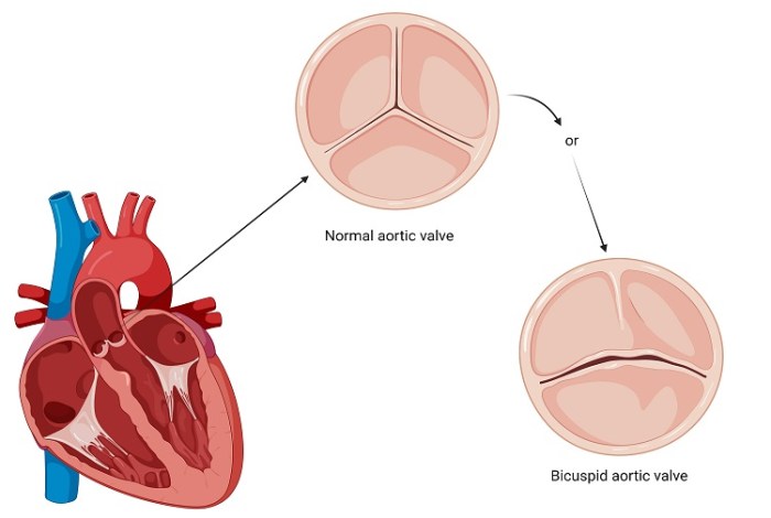
Bicuspid valve abnormalities are relatively common, affecting approximately 1-2% of the population. The most common abnormalities are stenosis and regurgitation.
- Stenosisoccurs when the valve leaflets become thickened and narrowed, obstructing blood flow from the left atrium to the left ventricle.
- Regurgitationoccurs when the valve leaflets do not close properly, allowing blood to leak back into the left atrium during ventricular systole.
Bicuspid valve abnormalities can lead to a variety of symptoms, including shortness of breath, chest pain, fatigue, and palpitations. Diagnosis of bicuspid valve abnormalities typically involves echocardiography and cardiac catheterization.
Surgical Intervention

Surgical intervention may be necessary to repair or replace a bicuspid valve that is causing significant symptoms. Valve repair involves repairing the existing valve leaflets and chordae tendineae. Valve replacement involves removing the existing valve and replacing it with a mechanical or biological valve.
Transcatheter aortic valve implantation (TAVI) is a less invasive procedure that can be used to replace a bicuspid valve in high-risk patients. TAVI involves inserting a new valve into the heart through a small incision in the leg.
Comparative Anatomy

The bicuspid valve is present in humans and other mammals. However, the structure and function of the bicuspid valve can vary between species.
- Mammals:In mammals, the bicuspid valve typically has two leaflets and is located between the left atrium and left ventricle.
- Birds:In birds, the bicuspid valve has only one leaflet and is located between the right atrium and right ventricle.
These differences in the structure and function of the bicuspid valve are likely due to evolutionary adaptations to different cardiovascular systems.
Expert Answers
What is the function of the bicuspid valve?
The bicuspid valve regulates blood flow from the left atrium to the left ventricle, ensuring proper circulation.
What are the symptoms of bicuspid valve abnormalities?
Symptoms may include shortness of breath, chest pain, fatigue, and palpitations.
How is a bicuspid valve abnormality diagnosed?
Echocardiography and cardiac catheterization are commonly used to evaluate valve function.